 |
|
 |
| |
 |
| Visiting Associate Professor |
|
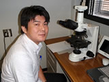
Shiro Miura
Department of Diagnostic Pathology Head (National Hospital Organization Nagasaki Medical Center)/
Visiting Associate Professor |
|
|
| Title & Licence : |
- Medical license (M.D.), 2003
- Doctor of Medicine (Ph.D) 2008
- Board Certified Pathologist of the Japanese Society of Pathology |
| Special field : |
- Human pathology
- Experimental Pathology
- Radiation life sciences |
Educational
background : |
- Nagasaki University School of Medicine
- Nagasaki University Graduate School of Biomedical Graduate School |
Work
experience : |
- Nagasaki University Hospital resident, 2002
- Researcher(postdoc), Nagasaki University Graduate School of Biomedical Sciences, 2008
- Assistant Professor, Graduate School of Biomedical Sciences, Nagasaki University, 2010
- Senior Assistant Professor, Atomic Bomb Disease Institute, Nagasaki University, 2014 |
Academic
Society : |
- The Japanese Society of Pathology
- The Japanese Society of Clinical Cytology
- The Japan Radiation Research Society
- Japan Endocrine Pathology Society |
| Awards: |
- 50th Japan Radiation Research Society Excellent Presentation Award |
My main research object is to understand the late health effect of radiation at molecular pathologic level. The incidence of several types of leukemia peaked during the 5 to 10 year period after the A-bomb explosions. Meanwhile, an increased risk of solid cancer has continued for decades, and the incidence of certain types of cancer still is higher than the incidence in controlled populations.
My recent study demonstrated the association of HER-2 and C-MYC oncogene amplification in breast cancers among A-bomb survivors with radiation exposure(Miura S, et al., Cancer 2008). Gene amplification is an important mechanism for oncogene overexpression in solid tumors and also serves as an indicator of genomic instability (GIN). Ionizing radiation effectively induces several DNA double-strand breaks (DSBs) in a dose-dependent manner, inducing a GIN. DSBs are repaired through error-prone, nonhomologous end joining; singlestrand annealing; and/or error-free, homologous recombination. Although most DNA damage is repaired correctly, it is well established that the repair process disrupts the genomic structure, which may manifest as induction of a mutation, gross rearrangement of chromatin, and promotion of tumorigenesis through the development of oncogene amplification. A-bomb radiation may induce minor disruptions of the genomic structures, which may result in GIN for an extended period in mammary glands and, subsequently, may affect oncogene amplification during breast carcinogenesis in the survivors.
I have established the tissue bank for solid cancers which were freshly resected from A-bomb survivors together with information on the A-bombing and medical data since April 2008. The establishment of A-bomb survivor’s tissue bank will enhance the global collaboration and contribute to understand the late health effects of radiation at molecular level, finding a key to the prediction, prevention and treatment of carcinogenesis as a late radiation effect. |
|
|
 |
|
Yuko Akazawa,
Professor (Department of Histology and Cell Biology) |
|
|
| Title & Licence : |
M.D., Ph.D.
Board Certified Member of the Japanese Society of Internal Medicine
Board Certified Gastroenterologist of the Japanese Society of Gastroenterology, Board Certified trainer
Board Certified Member of the Japan Gastroenterological Endoscopy Society
Board Certified Hepatologist of the Japan Society of Hepatology
Medical information manager
Board Certified Member of the Japan Gastroenterological Endoscopy Society, Board Certified trainer
|
| Special field : |
- Gastrointestinal medicine
|
Academic
Society : |
- The Japanese Society of Internal Medicine
- The Japanese Society of Gastroenterology (Councilor, Councilor of Kyusyu branch)
- Japan Gastroentelogical Endoscopy Society (Councilor of Kyusyu branch)
- The Japan Society of Hepatology
- Japan Society of Health Information Management
- The Japanese Society for Helicobactor Research (Representative)
- American Association for Study of Liver Diseases
- The Japanese Society of Pathology
- Japan Society of Histochemistry and Cytochemistry
- The Japanese Association of Anatomists (Representative)
- Japan Society for Cell Biology
|
 |
|
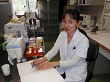
Mutsumi Matsuyama, Assistant Professor
(Division of Strategic Collaborative Research
Center for promotion of collaborative research on radiation and environment health effects) |
|
|
| Title & Licence : |
Ph.D. |
| Special field : |
- Experimental Pathology
- Radiation Effect Sciences
- Molecular pathology |
Educational
background : |
- Saga University, Department of Agriculture,
- Doctor of Medicine (Nagasaki-University) |
Work
experience : |
- GCOE Researcher (postdoc), Nagasaki University Graduate School of Biomedical Sciences, 2007-2012
- Assistant Professor, Graduate School of Biomedical Sciences, Nagasaki University, 2012- |
Academic
Society : |
- The Japanese Society of Pathology
- The Japan Radiation Research Society
- Japan Endocrine Pathology Society |
We have been researching for drugs that protect acute radiation damage in the intestinal tract after irradiation, especially radiation-induced cell death. It was reported that pretreatment of amino acid mixture, cystine and theanine improved the survival rate of rats after X-ray irradiation, and showed inhibition of apoptosis and increased proliferative activity of small intestinal crypt cells and bone marrow cells.
Exposure to ionizing radiation, especially during childhood, is a well-known risk factor for thyroid cancers. A large proportion of the thyroid cancer cases among children in the LSS cohort exposed to the bombs were attributed to radiation exposure, but the mechanism is not clear. To evaluate the radiosensitivity of immature and adult thyroid follicular epithelial cells in vivo, we analyzed the histological alterations, induction of apoptosis, cell proliferation, DNA damage response molecules, and induction of autophagy in rat thyroid glands in the acute phase and incidence of thyroid tumors in the chronic phase after external irradiation. In the acute phase, in both groups an increase in DNA damage response molecules was observed after irradiation, but cell death did not occur. A rapid decrease in the number of proliferating cells in young-aged rats with high proliferative activity, and an increased expression of autophagy-related molecule, LC3-II and p62 was observed. In the chronic phase, the incidence of thyroid tumors was high in young-aged rats, tumors occurred multiple, and abrogation of autophagy may be associated with radiation-induced thyroid carcinogenesis.
We are currently investigating how autophagy affects radiation-induced thyroid carcinogenesis in young-rats.
|
|
|
 |
| Senior Assistant Professor |
|
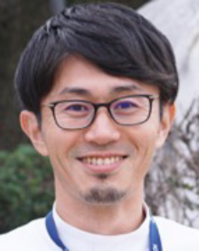
Ryota Otsubo, Senior Assistant Professor
(Department of Surgical Oncology) |
|
|
| Title & Licence : |
M.D., Ph.D.
Board Certified Surgeon of Japan Surgical Society, Board Certified trainer of Japan Surgical Society
Board Certified physician of Japanese Breast cancer Society, Board Certified trainer of Japanese Breast cancer Society
Board Certified Surgeon of Japan Association of Endocrine Surgery
Board Cancer Treatment Certified physician of Japanese Board of Cancer Therapy
Board certified physician of The Japan Central Organization on Quality Assurance of Breast Cancer Screening
Ministry of Health, Labor and Welfare certified clinical training instructor
JATEC・JPTEC instructor
|
| Special field : |
- Breast Surgery
- Endocrine Surgery |
Academic
Society : |
- Japan Surgical Society
- Japan Surgical Association
- Japanese Breast Cancer Society
- Japanese Association of Breast Cancer Screening
- Japanese Society of Thyroid Surgery
- The Japan Society of Human Genetics
- The Japanese Society of Pathology
- The Japanese Breast Cancer Society (Councilor)
- Member of Member Service Review Subcommittee, General Affairs Committee
- Member of the Committee for Revitalization of Local Regions (Kyushu Region)
- Member of the Committee for Evaluation of Clinical Guidelines
|
| (1) Kit and commercialization of a new novel diagnostic procedure for determining metastasis to sentinel lymph nodes in breast cancer using a semi-dry dot-blot (SDB) method.
Accurate assessment of metastasis of sentinel lymph nodes (SLNs) is important for deciding whether to remove axillary lymph nodes and providing appropriate treatment for patients. However, new diagnostic methods are required because there is a heavy workload for pathologists, and current methods have high false-negative rates and low sensitivity, and are expensive. Recently, we developed a “semi-dry dot-blot (SDB)” procedure, which visualizes the presence of cancer cells in the lavage fluid of sectioned lymph nodes by anti-pancytokeratin antibody (AE1/AE3) and chromogen on a dot-blot membrane. This method is based on the simple principle that there is usually no epithelial component in the lymph node if cancer does not develop into metastasis to the lymph node. We evaluated the validity and efficacy of the SDB method for the diagnosis of lymph node metastasis in a clinical setting.
To evaluate the efficacy of the SDB method in sentinel lymph node (SLN) biopsy, 174 SLNs from 100 cases of clinically node-negative breast cancer were analyzed. Each SLN was longitudinally sliced at 2-mm intervals and the sensitivity, specificity, accuracy, and time required for the SDB method were determined and compared with the permanent pathology report. Metastasis was detected in 15 SLNs (8.6%), and the sensitivity, specificity, accuracy, and mean required time of the SDB method were 93.3%, 96.9%, 96.6%, and 43.3 minutes, respectively. The cost for two SLN analyses by the SDB method was approximately 10 USD, which included cutting blades, primary and secondary antibodies, chromogen, filter, absorption paper, and dot-blot membrane. Special equipment was not necessary, but a centrifuge and micropipette were required.
The SDB method is simple, accurate, cost-effective, and feasible for the intraoperative diagnosis of SLN metastasis without the loss of lymph node tissue, and it enables a simultaneous pathological investigation and application to other cancers. We plan to develop a kit product, which will contribute to cost reduction for the detection of SLN metastasis. We succeeded in making this system as a rapid test kit, which is currently on clinical trial with support of Japan Agency for Medical Research and Development. We aim for future government approval.
(2) Preoperative diagnosis of thyroid follicular carcinoma using genomic instability
Thyroid follicular carcinomas are diagnosed by postoperative pathological diagnosis with views of capsule invasion, vascular invasion, or metastasis to other organ. Thus, it is difficult to diagnose preoperatively by fine-needle aspiration cytology. P53-binding protein 1 (53BP1) is one of nuclear proteins, that seems to reflect genomic instability of thyroid tumors. We already evaluated immunofluorescence analysis of 53BP1 expression in follicular carcinoma and follicular adenoma, and are trying to apply this result to the preoperative diagnosis of thyroid follicular tumors.
|
|
|
| |
| Nagasaki Harbor Medical Center |
|
Hiroyuki Yajima
Nagasaki Harbor Medical Center
(Department of Gastroenterology and Hepatology) |
|
|
| Title & Licence : |
M.D., Ph.D.
Board Certified Member of the Japanese Society of Internal Medicine, Board Certified Gastroenterologist of the Japanse Society of Gastroenterology, Board Certified Member of the Japan Gastroenterological Endoscopy Society |
| Special field : |
- Gastrointestinal medicine |
Academic
Society : |
- The Japanese Society of Internal Medicine
-
The Japanese Society of Gastroenterology
-
The Japan Gastroenterological Endoscopy Society
-
The Japanese Gastroenterological Association |
 |
|
Hideo Wada
Ureshino Medical Center(Department of Surgery) |
|
|
| Title & Licence : |
M.D., Ph.D.
Board Certified Surgeon of The Japanese Society of Gastrointestinal Surgery
Board Certified physician of Society of Gastrointestinal Surgery
Board Cancer Treatment Certified physician of Japanese Board of Cancer Therapy
Board Certified Surgeon of Japan Society for Endoscopic Surgery (General Surgery)
Board Certified Physician of Japanese Society of Abdominal Emergency Medicine
|
| Special field : |
- Gastroenterological Surgery |
Educational
background : |
- Graduation of Nagasaki University School of Medicine, 2005 |
Academic
Society : |
- Japan Surgical Society
- The Japanese Society of Gastroenterological Surgery
- Japan Surgical Association
- Japanese Society for Abdominal Emergency Medicine
- Japan Society for Endoscopic Surgery
- The Japanese Society of Gastroenterology |
| The molecular pathological analysis of neuroendocrine tumors in multiple organs |
|
|
| |
|
Kayoko Matsushima, Professor
(Medical Education Development Center) |
|
|
| Title & Licence : |
M.D., Ph.D.
Board Certified Member of the Japanese Society of Internal Medicine, Board Certified, Certified Instructor
Board Certified Gastroenterologist of the Japanese Society of Gastroenterology
Board Certified Member of the Japan Gastroenterological Endoscopy Society
Board Certified Member of the Japanese Gastroenterological Association of Gastroenterologist
Board Certified Hepatologist of the Japan Society of Hepatology
Board Certified Member of the Japanese Society for Helicobacter Research
Board Certified Member of the Japan Society for Oriental Medicine
|
| Special field : |
- Digestive disorders |
Academic
Society : |
- The Japanese Society of Internal Medicine
- The Japanese Society of Gastroenterology (Councilor of Kyusyu branch)
- Japan Gastroenterological Endoscopy Society (Councilor of Kyusyu branch)
- The Japan Society of Hepatology
- The Japanese Society for Helicobactor Research (Representative)
- The Japanese Gastroenterological Association
- Japan Society for Laser Surgery and Medicine (Councilor of Kansai branch)
- The Japan Society for Oriental Medicine (Councilor of Nagasaki Prefectural Committee)
- The Japan Society for Medical Education (Director, Representative)
|
 |
| Senior Assistant Professor |
|
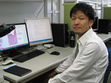
Keiichi Hashiguchi, Senior Assistant Professor
(Department of Endoscopy) |
|
|
| Title & Licence : |
M.D., Ph.D
Board Certified Member of the Japanese Society of Internal Medicine
Board Certified Gastroenterologist of the Japanese Society of Gastroenterology
Board Certified Member of the Japan Gastroenterological Endoscopy Society, Board Certified trainer
Board Certified gastroenterologist of the Japanese Gastroenterological Association, Board Certified trainer
|
| Special field : |
- Gastrointestinal medicine, Gastroenterological endoscope |
Academic
Society : |
- The Japanese Society of Internal Medicine
- The Japanese Society of Gastroenterology (Councilor of Kyusyu branch)
- Japan Gastroentelogical Endoscopy Society (Councilor of Kyusyu branch)
- The Japanese Gastroenterological Association
- The Japanese Society for Helicobactor Research Japanese Gastric Cancer Association
- The Japanese Society of Gastroenterology (Councilor of Kyusyu branch)
- Board Certified Member of the Japan Gastroenterological Endoscopy Society, Board Certified trainer (Councilor of Kyusyu branch, Academic councilor)
|
| Research contents |
 |
| Endoscopic diagnosis and treatment of superficial non-papillary duodenal epithelial tumors
Superficial non-ampullary duodenal epithelial tumors (SNADETs) are duodenal adenomas and adenocarcinomas arising from non-ampullary lesions. Recently, SNADETs have received increased recognition because of improvements in endoscopic devices and increased awareness among endoscopists. However, the guidelines for SNADETs have just been published and lack strong recommendations.
In endoscopic diagnosis, we are researching the ability to detect duodenal lesions by using Linked Color Imaging (Fujifilm Co., Ltd.), which is one of the image-enhanced endoscopies. If LCI mode is confirmed to be useful, it is expected that SNADETs can be detected and diagnosed by medical examination endoscopy.
Most SNADETs remain in the mucosa without lymph node metastasis; therefore, endoscopic treatment is often the first treatment choice. However, standardized endoscopic techniques to resect SNADETs are yet to be determined. Classical endoscopic mucosal resection (CEMR), a snare excision with submucosal injection, is commonly utilized for SNADETs <20 mm. Although CEMR is simple and safe, the anatomical characteristics of SNADETs often result in inadequate outcomes. For example, endoscopic maneuverability is often poor given their narrow lumen with sharp curves. Indeed, the en bloc resection and R0 resection rates of CEMR for SNADETs have been reported to be 63%–89% and 33.6%–62%, respectively. Lower R0 resection rates can cause local recurrence in up to 7.7% of cases. Following the introduction to the treatment of the stomach and colon, endoscopic submucosal dissection (ESD) has been introduced for SNADETs > 20 mm, and it has high en bloc and R0 resection rates (98.6%–100% and 86%–87.8%, respectively). However, duodenal ESD is a technically challenging procedure due to the extremely high risk of complications. In particular, the reported intraoperative and delayed perforation rates are 27.0%–35.7% and 6.3%–12.5%, respectively.
Underwater endoscopic mucosal resection (UEMR) has been recently introduced as an innovative endoscopic resection technique. The chief characteristic is water submersion to fill the duodenal lumen, instead of submucosal injection. Compared with CEMR, UEMR has the following advantages: lower possibility of unintentional snare fastening of the muscularis, ease of capture of relatively large lesions due to less duodenal lumen distension, and protection of the deeper duodenal wall from electrosurgical injury. In recent years, UEMR has become more widespread as a treatment for SNADETs < 20 mm. However, the reported negative horizontal margin and R0 resection rates remain moderate (46%–67%), resulting in a residual recurrence rate of 2.8%-5%.
Therefore, we designed "underwater EMR with submucosal injection and marking (UEMR-SIM) "as a modified combined method to improve the negative horizontal margin rate. UEMR-SIM has the following three underlying concepts: (i) underwater conditions for optimal snaring (the same as UEMR), (ii) marking that not only guides the lesion border in underwater conditions but also works as an anchor during snare deployment, and (iii) submucosal injection of a small volume of saline to expand the lesions and acquire additional horizontal margins.
We have started a prospective randomized controlled trial to investigate the efficacy of UEMR-SIM for SNADETs and compared with that of CEMR. If the efficacy of UEMR-SIM is proved, it is expected that a new endoscopic treatment for SNADETs will be established.
Establishment of a method for preventing delayed perforation of duodenal ESD using autologous myoblast sheets (joint research with the Department of Surgery, Tissue Engineering and Regenerative Therapeutics in Gastrointestinal Surgery, Nagasaki University Graduate School of Biomedical Sciences)
In duodenal ESD for SNADETs > 20 mm, the high rate of intraoperative and delayed perforation is problem. In particular, delayed perforation can lead to severe peritonitis and retroperitonitis. In recent years, laparoscopic and endoscopic cooperative surgery (LECS) is widespread for SNADETs. Although the closure of defect to prevent delayed perforation is suggested, there have not been established method.
We are conducting research to prevent delayed perforation by transplanting an autologous myoblast sheet in collaboration with Professor Kanetaka et al. The proof of concept has already been confirmed with a large animal experiment.
From April 2021, we have started a physician-led clinical trial "prevention of delayed perforation of duodenal ESD using an autologous myoblast sheet". If the usefulness of this regenerative medicine is proved, it is expected that the safety of endoscopic treatment for SNADETs will be developed.
|
|
|
 |
|
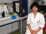
| Kazuko Shichijo, Visiting Researcher |
|
|
| Title & Licence : |
Philosophical Doctor (Medicine),
Pharmacist |
| Special field : |
- Experimental Pathology
- Human Pathology
- Molecular Pathology
- Radiation Science
- Pharmacology |
Educational
background : |
- Nagasaki University, Pharmaceutical Department
- Doctor of Medicine (Nagasaki-University) |
Work
experience : |
- Japan Society for the Promotion of Science; Research Associate, Duke University, Surgery Department, USA (Prof. N. Theodore N. Papas)
- Assistant Professor of Division of Tumor and Diagnostic Pathology, Atomic Bomb Institute, Nagasaki University Graduate School of Biomedical Sciences |
Academic
Society : |
- The Japanese Society of Ulcer Research (Councilor)
- The Japanese Pharmacological Society (Councilor)
- Japan Society of Neurovegetative Research (Councilor)
- The Japan Radiation Reseach Society
- The Japanese Society of Pathology |
| Lecture: |
- A lecturer in pathology at Nagasaki Women College |
| 1) |
Progress Research to health risk estimation, management and medical education of radiation
Plutonium remaining in the specimen of A-bomb victims at Nagasaki was determined by a classical method of autoradiography. These results may be a clue to investigate the effect of internally deposited radionuclide originated from A-bomb on human health, especially alpha-particle emitters with long half-lives. |
| 2) |
Applied research of regenerative medicine for tissue damage by high dose exposure to radiation
Radiation therapy causes radiation colitis and tumor. The extra-colonic cells contribute to abnormal repair in radiation colitis of rats transplanted with GFP marrow after sub-lethal irradiation. In the regenerative stage of radiation colitis, treatment of cytokine, DNA damage response and genomic instability in carcinogenesis were studied. |
| 3) |
Collaboration with Hiroshima University, H24“Investigation of plutonium and internal exposure in A-bomb victims-part 2”
Alpha-particle emitters remaining in the specimen of A-bomb victims at Nagasaki was determined. |
|
|
 |
|
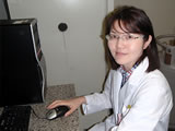
Tomomi Kurashige, Visiting Researcher
(Department of Molecular Medicine) |
|
|
| Title & Licence : |
Ph.D. |
| Special field : |
- Experimental pathology
- Radiation life sciences |
Academic
Society : |
- Japan Endocrine Pathology Society
- Japanese Cancer Association |
| Current research topic |
 |
| 1) |
Comparative study of energy metabolism between oncocytoma and non-oncocytoma of the thyroid gland and its application to treatment
Thyroid oncocytoma is a rare thyroid tumor in which abnormal mitochondria, in which the electron transport chain complex I does not function due to abnormal mitochondrial DNA (mDNA), are accumulated in the cytoplasm. Since this type of tumor cell cannot produce ATP by oxidative phosphorylation in mitochondria, it is considered that ATP production depends on substrate level phosphorylation in glycolysis and TCA cycle. Therefore, thyroid oncocytoma is expected to be more susceptible to cytotoxicity due to inhibition of glycolysis and inhibition of glutamine metabolism, which is important for substrate level phosphorylation in the TCA cycle than for typical thyroid cancer. Therefore, we are conducting research to compare the susceptibility of thyroid oncocytoma to typical thyroid cancer cell lines to various metabolic inhibitors and to identify the predominant ATP production pathway in each. We are also investigating the involvement of mitochondrial function by creating a mitochondrial DNA-deficient cells (ρ0). Furthermore, we plan to investigate the combined enhancement effect of these metabolism inhibitors and conventional thyroid cancer treatment methods (radiation therapy, kinase inhibitors, etc.). |
| 2) |
Elucidation of radiation-induced thyroid carcinogenic mechanism using thyroid-specific ATG5KO mice
We are conducting research to clarify the role of autophagy in the thyroid gland and whether autophagy suppresses or promotes carcinogenesis when thyroid exposed to external radiation. Specifically, we are observing changes in autophagy when growth stimulation / suppression is given to normal thyroid gland, and investigating thyroid function at promotion / suppression of drug-induced autophagy. Furthermore, by observing Atg5TPO-KO mice for a long period of time, we are investigating whether autophagy deficiency influences thyroid function and morphological changes, as well as the frequency of carcinogenesis due to irradiation and the involvement of ROS production. |
|
|
 |
|
Luong Thi My Hanh,
Postdoctoral Researcher, Karolinska Institutet |
|
|
| Title & Licence : |
M.D., Ph.D.
M.D. (Hanoi Medical University) |
| Special field : |
- Human pathology |
Academic
Society : |
- The Japanese Society of Pathology |
|
|

