Leukemia
A: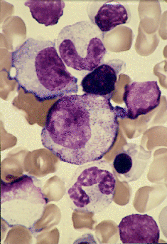 B:
B: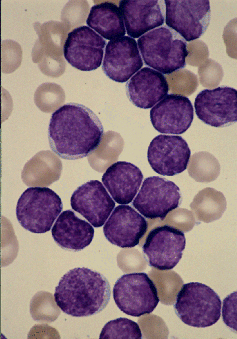
C: D:
D: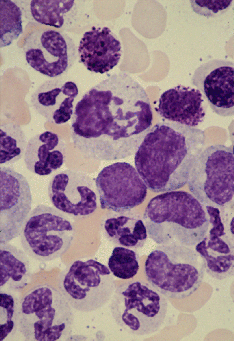
A : Picture of bone marrow smear (control); Normal granulocytes and
erythroblasts are evident.
B : Acute lymphoid leukemia (ALL); There is a marked proliferation of
small lymphoblasts.
C : Acute myeloid leukemia (AML); There is a marked proliferation of
large myeloblasts.
D : Chronic myeloid leukemia (CML); There is a marked proliferation of
granulocytes at various stages of maturation.
E : Crude incidence graph (166KB gif)
- Unlike solid tumors, radiation-induced leukemia occurred from
around two years after the atomic bombing. Both the absolute and
relative risk reached a peak shortly thereafter and then gradually
decreased.
- A dose response relationship was recognized in acute lymphoid
leukemia (ALL), acute myeloid leukemia (AML) and chronic myeloid
leukemia (CML).
- The risk of leukemia was high in the young age group ATB.
- Adult T-cell leukemia is an endemic disease in Nagasaki and did
not show a dose response relationship.
- CML is more frequent in Hiroshima than in Nagasaki because the
spontaneous incidence of the disease is three times higher in the
former than in the latter, and no significant difference was observed
in the excess incidence of radiation-induced CML between the two cities.
Return to INDEX
Data Distributed from
Scientific Data Center for the Atomic Bomb Disaster,
Nagasaki University.
HTML Created by datactr_a_bomb@ml.nagasaki-u.ac.jp
 B:
B:
 D:
D: